Van Gieson Stain Liver: Understanding the Histological Analysis
Van Gieson stain liver is a crucial aspect of histological analysis, providing valuable insights into the liver's structural and functional properties. This stain is specifically designed to highlight the collagen fibers in liver tissue, allowing researchers and medical professionals to assess liver health and diagnose potential conditions. With its widespread application in medical research and diagnosis, understanding van Gieson stain liver is essential for experienced hobbyists and professionals alike.
What is Van Gieson Stain?
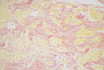
Van Gieson stain is a histological staining technique used to visualize collagen fibers in tissue samples. Developed by van Gieson, this stain combines picric acid and acid fuchsin to produce a distinctive red coloration of collagen fibers, while other tissue components appear yellow or brown. This technique has been widely adopted in medical research and diagnosis, particularly in the analysis of liver tissue.
The van Gieson stain is particularly useful in assessing liver health, as it allows researchers to examine the distribution and density of collagen fibers in liver tissue. This information can be used to diagnose a range of liver conditions, including fibrosis and cirrhosis.

How Does Van Gieson Stain Work in Liver Tissue?
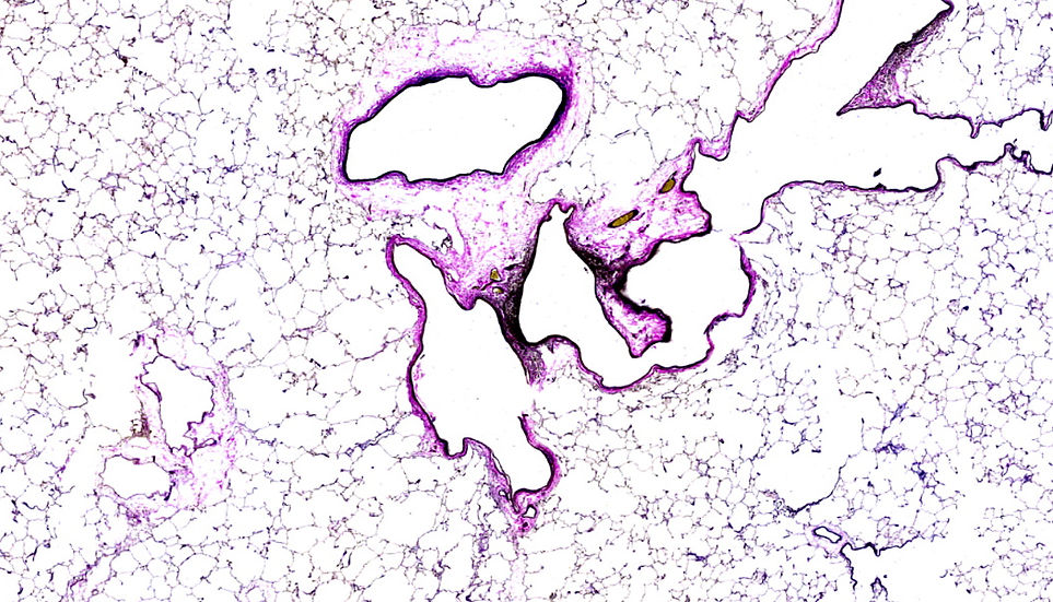
The van Gieson stain works by binding to collagen fibers in liver tissue, producing a distinctive red coloration. This allows researchers to visualize the distribution and density of collagen fibers, which can be indicative of liver health. In healthy liver tissue, collagen fibers are typically sparse and evenly distributed, whereas in diseased tissue, collagen fibers may be more abundant and irregularly distributed.
The van Gieson stain can be used in conjunction with other staining techniques to provide a more comprehensive understanding of liver tissue. For example, the stain can be used in combination with hematoxylin and eosin (H&E) staining to provide a more detailed understanding of liver morphology. 
What are the Implications of Van Gieson Stain Liver Analysis?
The analysis of van Gieson stain liver tissue has significant implications for our understanding of liver health and disease. By providing a detailed understanding of collagen fiber distribution and density, the van Gieson stain can be used to diagnose a range of liver conditions, including fibrosis and cirrhosis. Additionally, the stain can be used to monitor the progression of liver disease and assess the effectiveness of treatments.
In conclusion, van Gieson stain liver is a critical aspect of histological analysis, providing valuable insights into the structural and functional properties of liver tissue. By understanding the principles and applications of the van Gieson stain, experienced hobbyists and professionals can gain a deeper appreciation for the complexities of liver health and disease. As research continues to uncover the intricacies of liver function and disease, the van Gieson stain is likely to remain a vital tool in the diagnosis and treatment of liver conditions.
Verhoeff Van Gieson Stain | Elastic & Collagen Fiber Detection | IHisto
Verhoeff Van Gieson Stain | Elastic & Collagen Fiber Detection | iHisto
Celnovte
Celnovte
D: Cirrhotic Liver, Van Gieson Technique. | Download Scientific Diagram

d: Cirrhotic liver, van Gieson technique. | Download Scientific Diagram
Liver Biopsy With Haematoxylin/van Gieson Stain Showing Periportal

Liver biopsy with haematoxylin/van Gieson stain showing periportal ...